
Key Takeaways
- OCT provides a 3D cross-section of the retina, ideal for spotting swelling or holes.
- Fundus Photography acts like a digital map of the back of the eye.
- Fluorescein Angiography uses dye to find leaks in blood vessels.
- OCT Angiography (OCTA) is a newer, dye-free way to see blood flow.
- Combining these tools (multimodal imaging) gives the most accurate diagnosis for diseases like diabetic retinopathy.
The Digital Map: Fundus Photography
Think of Fundus Photography as a high-resolution photo of the back of your eye. The "fundus" is simply the interior surface of the eye opposite the lens. Using specialized cameras-such as the Zeiss FF 450+-doctors take a 2D image of the posterior pole, including the macula, the optic disc, and the retinal vessels.
If you've ever had a photo taken where a bright flash momentarily blinded you, that's likely a fundus photo. It doesn't show depth, but it's incredibly useful for documenting the overall health of the retina over time. For example, if a doctor sees a new hemorrhage or a change in the color of the optic nerve, they have a baseline image to compare it to. It's the first step in most retinal evaluations because it tells the doctor where to look more closely with other tools.
Seeing in 3D: Optical Coherence Tomography (OCT)
While a photo is 2D, Optical Coherence Tomography (or OCT) is more like an ultrasound, but it uses light instead of sound. It creates cross-sectional images, allowing doctors to see the different layers of the retina as if they had sliced it open and looked at it from the side.
Modern clinics mostly use Spectral-Domain OCT (SD-OCT), which provides an axial resolution of about 5-7 micrometers. This level of detail is so precise that doctors can spot microscopic pockets of fluid or "holes" in the macula that would be invisible on a standard photo. For those with more complex needs, Swept-Source OCT (SS-OCT) offers even deeper penetration into the choroid (the layer of blood vessels beneath the retina), performing up to 400,000 A-scans per second.
OCT is a game-changer for conditions like age-related macular degeneration. Instead of guessing if a treatment is working, a doctor can literally measure the thickness of the retinal layers in microns and see the fluid disappearing in real-time.
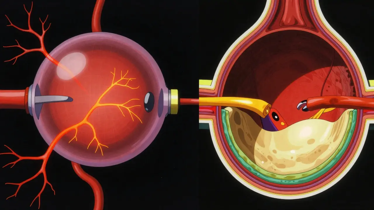
Tracing Blood Flow: Fluorescein Angiography
Sometimes, looking at the structure isn't enough; doctors need to see how the blood is actually moving. This is where Fluorescein Angiography (FA) comes in. Unlike the previous tests, this one is invasive. A bright orange dye called fluorescein is injected into a vein in your arm, and as it travels to the eye, a series of rapid-fire photos are taken.
Why go through the trouble of an injection? Because dye "leaks" where vessels are damaged. In cases of diabetic macular edema, FA is often more sensitive than OCT for detecting low-grade leakage. If a blood vessel is slightly "leaky," the dye will pool in that area, creating a bright spot that tells the doctor exactly where the pathology is located. It's the gold standard for visualizing the dynamic flow of blood and identifying areas where the retina isn't getting enough oxygen (non-perfusion).
| Feature | Fundus Photography | OCT / SD-OCT | Fluorescein Angiography | OCT Angiography (OCTA) |
|---|---|---|---|---|
| Invasiveness | Non-invasive | Non-invasive | Invasive (Dye injection) | Non-invasive |
| Image Type | 2D Surface Map | 3D Cross-section | Blood Flow/Leakage | 3D Vascular Map |
| Time Required | Seconds | Minutes | 10-30 Minutes | Seconds |
| Best For... | General Screening | Retinal Thickness/Fluid | Vessel Leakage | Capillary Perfusion |
The New Frontier: OCT Angiography (OCTA)
In recent years, a breakthrough called OCT Angiography (or OCTA) has started to replace dye-based tests. OCTA uses the motion of red blood cells to create a map of the vasculature without needing any dye. It's fast, safe, and gives a three-dimensional view of the superficial, middle, and deep capillary plexuses of the fovea.
For patients, the benefit is obvious: no needles and no risk of allergic reactions to fluorescein. For doctors, OCTA is superior for spotting "neovessels" (abnormal new blood vessels) in proliferative diabetic retinopathy. It can find tiny areas of non-perfusion-spots where blood isn't flowing-that are too small to be seen on a traditional FA scan. For instance, in conditions like punctate inner choroidopathy (PIC), OCTA can detect non-perfusion in the choriocapillaris that other imaging simply misses.
However, it's not perfect. OCTA can't see "leakage." It shows where the vessels are, but it can't tell you if those vessels are leaking fluid into the retina. That's why doctors often use a "multimodal" approach, combining OCTA with traditional angiography to get the full picture.
Putting it Together: Multimodal Imaging in Practice
No single test provides all the answers. A doctor treating a patient with Coats disease, for example, will use several of these tools. They might use fundus photos to see the overall extent of the disease, then an OCT to find exudates (leaky fats) hidden in multiple retinal layers, and finally an FA to see the abnormal vascular patterns.
This layered approach is critical because different diseases hide in different ways. Macular holes are best spotted with SD-OCT, while severe retinal vein occlusions often require FA to determine the severity of the edema. By layering a 2D map (Fundus), a 3D structure (OCT), and a flow analysis (FA or OCTA), the physician creates a comprehensive diagnostic profile.
The only real downside to these high-tech scans is the requirement for patient cooperation. OCT and OCTA are very sensitive to movement. If a patient blinks or shifts their gaze, it can create "motion artifacts"-blurry lines that can be mistaken for vascular abnormalities. This makes the tests challenging for children or patients with advanced dementia who struggle to keep their eyes fixed on the target.
Does the dye in angiography stay in my system?
Fluorescein dye is temporary. It is filtered by the kidneys and typically leaves your system within 24 hours. You might notice your skin or urine has a yellow-orange tint for a short period after the procedure, which is completely normal.
Is OCT better than a fundus photo?
It's not about being "better," but about providing different information. A fundus photo is a surface view (like a map of a city), while OCT is a cross-section (like looking at the blueprints of a building's floors). You need the photo to see the overall layout and the OCT to see the internal damage.
Can OCTA completely replace fluorescein angiography?
Not yet. While OCTA is faster and safer, it cannot detect "leakage." If a doctor needs to know if a vessel is leaking fluid into the macula, they still need the dye-based fluorescein angiography to confirm it.
Are there any risks associated with these imaging tests?
OCT and fundus photography are non-invasive and have virtually no risk. Fluorescein angiography carries a small risk of allergic reaction to the dye, which is why clinics ask about allergies beforehand. Rare but serious reactions can occur, though they are manageable in a clinical setting.
How often do I need these scans?
This depends on your condition. For a healthy eye, a fundus photo every few years might suffice. However, for someone with active macular degeneration or diabetic retinopathy, OCT scans may be performed every few weeks or months to monitor the effectiveness of injections or laser treatments.
Next Steps and Troubleshooting
If you are scheduled for an imaging appointment, here are a few tips to ensure the best results:
- Dilation: Be prepared for your pupils to be dilated. This allows the cameras to see the back of the eye clearly. Bring sunglasses, as the world will be very bright afterward.
- Stability: During OCT or OCTA, you'll be asked to look at a target. Try to keep your eye as still as possible. If you feel like you're drifting, let the technician know so they can help reposition you.
- Dye Prep: If you're getting a fluorescein angiography, make sure to disclose all allergies and current medications. Stay hydrated, as this helps your kidneys clear the dye more efficiently.
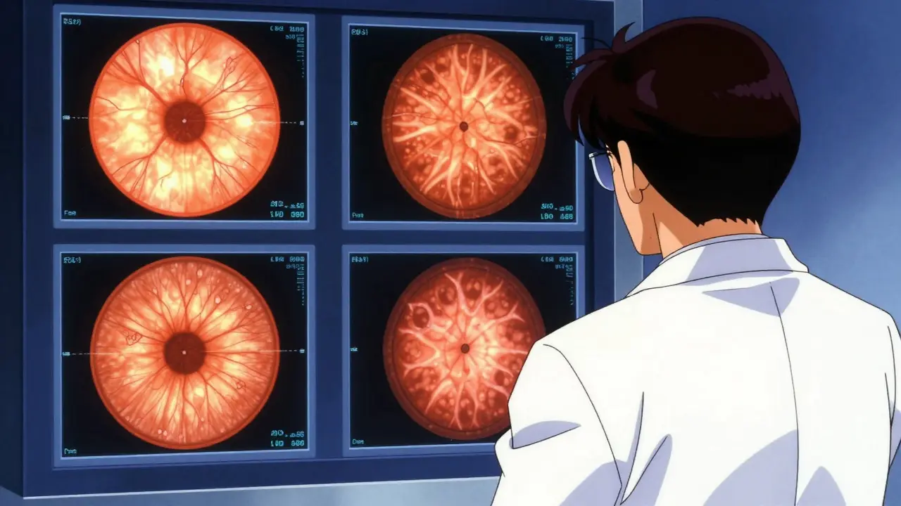
- Results: Don't panic if the technician doesn't give you an immediate answer. These images require expert interpretation by an ophthalmologist to distinguish between true pathology and imaging artifacts.
Daniel Runion
April 27, 2026 AT 04:43Imagine thinking a simple table is enough to explain these complex systems!!! Honestly, the oversimplification here is just... tragic. I've seen far more detailed breakdowns in a basic textbook, and the lack of mention regarding the specific laser wavelengths used in SD-OCT is a glaring omission... simply unacceptable!!!
William Zhigaylo
April 28, 2026 AT 06:37The audacity of presenting this as a comprehensive guide is staggering. It is fundamentally irresponsible to gloss over the systemic risks of fluorescein angiography in patients with renal impairment. You have provided a superficial overview that borders on negligence, failing to emphasize the severe psychological distress associated with invasive ocular diagnostics.
Ben Jima
April 28, 2026 AT 17:10Great breakdown of the tools! For anyone getting an OCT, just remember to take a deep breath and relax your shoulders; it actually helps you keep your eye steady on the target and makes the technician's job much easier. You've got this!
Brittney Prince
April 29, 2026 AT 06:31Sure, it's a "diagnostic tool" but why are they so obsessed with mapping the inside of our eyes? It feels like they're just collecting biometric data for some massive database we aren't told about. The dye is probably just a cover for something else they're injecting into us.
Anand Mehra
May 1, 2026 AT 04:34imaging is just a mirror of the void. tools change but the blindness stays the same. basically just paying for fancy photos of a failing organ
Nila Sawyer
May 2, 2026 AT 12:28Oh my goodness, I just love how this explains everything so clearly for people who might be scared of the doctors office! 🌟 It is so important to stay positive and informed about our health because once we understand the science behind these amazing machines, the anxiety just melts away and we can focus on the wonderful gift of sight! ✨ Keep sharing this kind of knowledge because it truly empowers us all to take charge of our wellness journey with a smile on our faces! ❤️🌈
Nikita Shabanov
May 3, 2026 AT 01:20To add a bit more context for those wondering about the "motion artifacts" mentioned, this is particularly common in patients with nystagmus. In those cases, clinicians might use specialized software to digitally stabilize the image or perform the scan more frequently to find a usable frame. It's a common hurdle in retinal imaging but manageable with a patient technician.